Blood Collection Tubes Utilized for Fasting Blood Sugar Measurement
A Source of Variation in Clinical Laboratory Test Results
DOI:
https://doi.org/10.21141/PJP.2026.601Keywords:
Clinical Laboratory Science, Blood Collection Tubes, Diabetes mellitus, Fasting Blood SugarAbstract
Background. Diabetes mellitus (DM) remains a major global health burden, with increasing prevalence in developing countries such as the Philippines. Accuracy of glucose measurement is vital for diagnosis and management; however, preanalytical variables, particularly glycolysis from varying collection tubes, temperature and time interval can significantly affect test results. Despite guideline recommendations favoring plasma, serum is still commonly used in local clinical practice.
Objective. This study evaluated the effectiveness of different commercially available blood collection tubes in preserving glucose stability by minimizing pre-analytical glycolysis.
Methodology. A cross-sectional observational and quasi-experimental study was conducted among 40 healthy adult participants (18–59 years) from a tertiary institution in Quezon City, Philippines. A total of 160 samples were collected using four tube types (one plasma and three serum tubes). Samples were analyzed at varying time intervals (0–180 minutes) and storage conditions (room temperature and 4°C). Glucose levels were measured using the glucose oxidase method. Statistical analysis included Shapiro–Wilk, Kruskal–Wallis H-test, Welch’s t-test, and Dwass–Steel–Critchlow–Fligner post hoc comparisons at a 1% significance level.
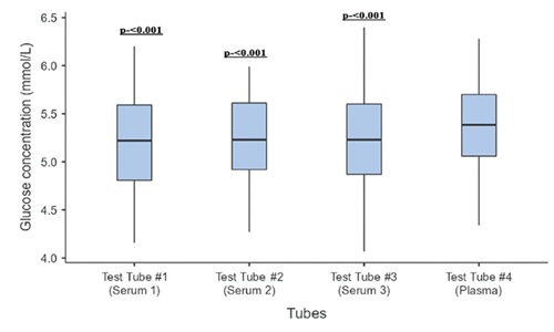
Results. Glucose concentrations differed significantly across tube types (p <0.001) and time intervals (p <0.001), but not by storage temperature (p = 0.023). Plasma samples demonstrated significantly higher glucose levels than serum samples, with all serum tubes showing a consistent negative bias relative to the fluoride-containing plasma tube. Although differences remained within the ± 6 mg/dL CLIA allowable total error, clinically relevant deviations were observed near diagnostic thresholds. Glucose stability significantly declined beyond 60 minutes, indicating ongoing glycolysis despite sample separation.
Conclusion. Blood collection tube type and delayed processing significantly influence glucose measurements. While acceptable within analytical limits, systematic biases may affect clinical interpretation. Standardization of blood collection practices and stricter pre-analytical protocols are essential to improve diagnostic accuracy for diabetes in the Philippines.
Downloads
References
Bishop ML, Fody EP, Schoeff LE. Clinical Chemistry, 8th ed. Wolters Kluwer; 2018.
Yang C, Feng W, Li Y, Tian X, Zhou Z, Lu L, Nie Y. A promising method for diabetes early diagnosis via sensitive detection of urine glucose by Fe-Pd/rGO. Dyes Pigments. 2019;164:20-6. https://doi.org/10.1016/j.dyepig.2018.12.061
Ng JYS, Clement IJ, Jimeno C, et al. Estimating direct medical costs of type 2 diabetes mellitus in the Philippines: a protocol. BMJ Open. 2020;10(7):e025696. https://pubmed.ncbi.nlm.nih.gov/32723733 https://www.ncbi.nlm.nih.gov/pmc/articles/PMC7389482 https://doi.org/10.1136/bmjopen-2018-025696
Cando LFT, Quebral EPB, Ong EP, et al. Current
status of diabetes mellitus care and management in the
Philippines. Diabetes Metab Syndr. 2024;18(2):102951.
https://pubmed.ncbi.nlm.nih.gov/38382166 https://doi.org/10.1016/j.dsx.2024.102951
American Diabetes Association. Classification and diagnosis of diabetes: standards of medical care in diabetes-2021. Diabetes Care. 2021;44(Suppl 1):S15-33. https://pubmed.ncbi.nlm.nih.gov/33298413 https://doi.org/10.2337/dc21-S002
McPherson RA, Pincus MR, eds. Henry's clinical diagnosis and management by laboratory methods. 24th ed. Elsevier; 2022. ISBN: 9780323673204
Kubihal S, Goyal A, Gupta Y, Khadgawat R. Glucose measurement in body fluids: a ready reckoner for clinicians. Diabetes Metab Syndr. 2021;15(1):45-54. https://pubmed.ncbi.nlm.nih.gov/33310176 https://doi.org/10.1016/j.dsx.2020.11.021
Landgraf R, Nauck M, Freckmann G, et al. Pitfalls in the diagnosis of diabetes: are we too lax with laboratory parameters? Dtsch Med Wochenschr. 2018;143(21):1549-55. https://pubmed.ncbi.nlm.nih.gov/30235490 https://doi.org/10.1055/a-0673-2156
Lee H, Hong YJ, Baik S, Hyeon T, Kim DH. Enzyme-based glucose sensor: from invasive to wearable device. Adv Healthc Mater. 2018;7(8):e1701150. https://pubmed.ncbi.nlm.nih.gov/29334198 https://doi.org/10.1002/adhm.201701150
Lippi G, Nybo M, Cadamuro J, Guimaraes JT, van Dongen-Lases E, Simundic AM. Blood glucose determination: effect of tube additives. Adv Clin Chem. 2018;84:101-23. https://pubmed.ncbi.nlm.nih.gov/29478513 https://doi.org/10.1016/bs.acc.2017.12.003
Bonetti G, Carta M, Lapolla A, Miccoli R, Testa R, Mosca A; SIBioC-SIPMeL Working Group on Diabetes and the Italian Diabetes Society (SID). Correct determination of glycemia in the diagnosis and management of diabetes: recommendations for the optimization of the pre-analytical phase. Nutr Metab Cardiovasc Dis. 2019;29(1):1-3. https://pubmed.ncbi.nlm.nih.gov/30482424 https://doi.org/10.1016/j.numecd.2018.09.013
Winter T, Hannemann A, Suchsland J, Nauck M, Petersmann A. Long-term stability of glucose: glycolysis inhibitor vs gel barrier tubes. Clin Chem Lab Med. 2018;56(8):1251-8. https://pubmed.ncbi.nlm.nih.gov/29525788 https://doi.org/10.1515/cclm-2017-0860
Pan CT, Francisco MD, Yen CK, Wang SY, Shiue YL. Vein pattern locating technology for cannulation: a review of the low-cost vein finder prototypes utilizing near infrared (NIR) light to improve peripheral subcutaneous vein selection for phlebotomy. Sensors (Basel). 2019;19(16):3573. https://pubmed.ncbi.nlm.nih.gov/31426370 https://www.ncbi.nlm.nih.gov/pmc/articles/PMC6719195 https://doi.org/10.3390/s19163573
Francisco MD, Chen WF, Pan CT, et al. Competitive real-time near infrared vein finder imaging device to improve peripheral subcutaneous vein selection in venipuncture for clinical laboratory testing. Micromachines (Basel). 2021;12(4):373. https://pubmed.ncbi.nlm.nih.gov/33808493 https://www.ncbi.nlm.nih.gov/pmc/articles/PMC8067297 https://doi.org/10.3390/mi12040373
Leung GKW, Huggins CE, Ware RS, Bonham MP. Time of day difference in postprandial glucose and insulin responses: systematic review and meta-analysis of acute postprandial studies. Chronobiol Int. 2020;37(3):311-26. https://pubmed.ncbi.nlm.nih.gov/31782659 https://doi.org/10.1080/07420528.2019.1683856
Pant V, Gautam K, Pradhan S, Pyakurel D, Shrestha A. Blood glucose concentration measured in EDTA/F plasma and serum in a referral clinical laboratory in Nepal. J Pathol Nepal. 2021;11(1):1837-41. https://doi.org/10.3126/jpn.v10i2.32351
Serin Y, Acar Tek N. Effect of circadian rhythm on metabolic processes and the regulation of energy balance. Ann Nutr Metab. 2019;74(4):322-30. https://pubmed.ncbi.nlm.nih.gov/31013492 https://doi.org/10.1159/000500071
Bazzano G, Galazzi A, Giusti GD, Panigada M, Laquintana D. The order of draw during blood collection: a systematic literature review. Int J Environ Res Public Health. 2021;18(4):1568. https://pubmed.ncbi.nlm.nih.gov/33562241 https://www.ncbi.nlm.nih.gov/pmc/articles/PMC7915193 https://doi.org/10.3390/ijerph18041568
McCall RE. Phlebotomy essentials, 8th ed. Jones and Bartlett Learning; 2021.
Keohane EM, Otto CN, Walenga JM. Rodak's Hematology: Clinical principles and applications. 6th ed. Elsevier; 2020.
Burtis CA, Ashwood ER, Tietz NW. Tietz fundamentals of clinical chemistry and molecular diagnostics, 7th ed. Elsevier; 2018.
Ayer T, Zhang C, Zeng C, White CC, Joseph VR. Analysis and improvement of blood collection operations. Manuf Serv Oper Manag. 2019;21(1):29-46. https://doi.org/10.1287/msom.2017.0693
Chadwick K, Whitehead SJ, Ford C, Gama R. kEDTA sample contamination: a reappraisal. J Appl Lab Med. 2019;3(6):925-35. https://pubmed.ncbi.nlm.nih.gov/31639684 https://doi.org/10.1373/jalm.2018.027920
Becton Dickinson and Company. BD vacutainer evacuated blood collection system; 2020. https://www.bd.com/documents/guides/directions-for-use/PAS_BC_BD-Vacutainer-Evacuated-Blood-Collection-System_SF_EN.pdf
Dickson LM, Buchmann EJ, Janse Van Rensburg C, Norris SA. The impact of differences in plasma glucose between glucose oxidase and hexokinase methods on estimated gestational diabetes mellitus prevalence. Sci Rep. 2019;9(1):7238. https://pubmed.ncbi.nlm.nih.gov/31076622 https://www.ncbi.nlm.nih.gov/pmc/articles/PMC6510785 https://doi.org/10.1038/s41598-019-43665-x
Loganathan P, Gasper SK, Afel FK, Kandaswamy S. Pre-analytical errors in glucose estimation results in query on diabetic management. Indian J Clin Biochem. 2020;35(1):32-42. https://pubmed.ncbi.nlm.nih.gov/32071494 https://www.ncbi.nlm.nih.gov/pmc/articles/PMC6995467 https://doi.org/10.1007/s12291-018-0782-6
Pleus S, Freckmann G, Baumstark A, Haug C. Stability of glucose concentrations in frozen plasma. J Diabetes Sci Technol. 2021;15(3):745-51. https://doi.org/10.1177/1932296820963657
Bhargava M, Singh NP, Gupta AK. Should we still collect blood glucose sampling in fluoride tubes? An evidence-based study. Int J Diabetes Dev Ctries. 2019;39(2):243-4. https://doi.org/10.1007/s13410-018-0688-0
Bhatt MP, Shrestha S. Rate of glucose utilization by blood cells in serum and plasma specimens with or without using preservative. Mod Med Lab J. 2022;17(5):1-9. https://doi.org/10.30699/mmlj17.5.1.1
Downloads
Published
How to Cite
Issue
Section
Categories
License
Copyright (c) 2026 Jomar Adams Ganding

This work is licensed under a Creative Commons Attribution-NonCommercial-ShareAlike 4.0 International License.


 @philippinepathologyjournal